 0866 022 097
 institute@mescells.com
Contamination Control in the Cell Culture Room: Rigorous Procedures That Deliver Quality and Safety


In the microscopic world of biotechnology, an invisible mistake can lead to visible and costly failures. The risk of contamination in the laboratory is not just a technical risk; it is a constant, direct threat to the accuracy of research data, the safety of patients and the reputation of biomedical organizations.
For scientists, a contaminated culture dish means months of work down the drain; for patients, it means the risk of dangerous complications when cells are introduced into the body. Recognizing this, contamination control in the culture room has been elevated to an art that combines human discipline with modern technology. In the article below, Mescells helps you better understand contamination risks in the culture room and how technology is applied.
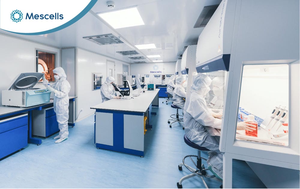
Scientists work under strict procedures to ensure contamination control in an international-standard culture room
1. Why is contamination control needed in the culture room?
Cell culture is, at its core, a feat of biotechnology that recreates life outside the body. As defined by biomedical experts, it is the process of isolating cells from the tissue of a multicellular organism and maintaining their growth in an artificial environment in which nutrients, temperature and gases are strictly controlled. When supplied with the right growth factors, these cells behave as independent units, able to divide and carry out their characteristic biological functions.
Contamination control in the culture room is central to maintaining a stable environment and protecting the target cell population, because of the enormous biological gap between animal cells and microorganisms. While animal cells are “demanding guests” that need 18-24 hours to double once, foreign bacteria can double their population every 30 minutes. If a sterile barrier is breached, bacteria quickly take over, consume all the nutrients and alter the pH of the medium, leading to the death of the target cell population.

Every medium supplementation step requires strict contamination control in the culture room to keep cells alive
Today, major research centers use two main culture methods:
- 2D culture: Cells are grown on flat plastic surfaces (Petri dishes or flasks), adhering as a monolayer. This is the foundational method and makes morphological changes easy to observe.
- 3D culture (bioreactors): This is a major step forward, allowing cells to grow in three-dimensional space and form spheroids that mimic the structure of real tissue in the body (in vivo).
Maintaining a sterile environment is not just about keeping cells alive. More importantly, it ensures the stability of the phenotype and genotype. A contaminated environment triggers cellular stress responses, leading to unwanted “transformation”. Cells may become immortal or carry mutations that risk causing tumors when transplanted into humans. Contamination control is therefore the “insurance” for the clinical value of stem cells.

Diagram of 2D mesenchymal stem cell culture with contamination control points
2. Contaminants that must be controlled in the culture room
In the laboratory, contamination control in the culture room requires technicians to recognize contaminants and signs of contamination early, because many risks cannot be detected with the naked eye at the outset.
2.1. Bacterial contamination
This is the most common and most easily recognized type of contamination. Just a few bacterial cells getting in will turn the culture medium from clear to cloudy (turbidity) overnight. Under the microscope, small rod-shaped or spherical particles can be seen moving chaotically. The pH change caused by bacterial metabolism usually turns the medium (which contains the phenol red indicator) yellow, accompanied by a characteristic foul smell.
2.2. Fungal and yeast contamination
Molds usually enter through the air and grow quickly when culture conditions are favorable. The typical sign is the appearance of filaments (hyphae) that look like small white cotton tufts floating on the surface of the medium. Yeasts are more discreet: they are usually oval, reproduce by budding and form refractile particles that gather in clusters, making the medium slightly cloudy while the pH changes more slowly than with bacteria.

Mold hyphae grow into a dense network that looks like spreading white cotton tufts
2.3. Mycoplasma and the HeLa cell line
The most dangerous of these agents is Mycoplasma. This bacterium has no cell wall, is extremely small (0.15-0.3 μm), does not cloud the medium and cannot be seen under an ordinary light microscope. Yet it silently alters the metabolism and gene expression of host cells. Statistics from specialist lectures by Prof. Ahmed Ali Mohammed indicate that as many as 15-20% of experiments worldwide are skewed by Mycoplasma contamination or cross-contamination between cell lines.

Visual comparison of a clean culture flask and a cloudy contaminated dish
Cross-contamination in particular is a frightening risk. The HeLa cell line, an immortal cancer cell line that grows extremely vigorously, is often regarded as the “villain” of this story. Just a few HeLa cells on a pipette can invade and destroy your precious stem cell line, rendering the research results worthless.
Comparison of the main types of contamination:
| Type of contamination | Signs | Growth rate | Effect on cells |
| Bacteria | Cloudy medium, falling pH (yellow), odor | Very fast (doubling every 30 minutes) | Kills cells quickly |
| Mold | White fuzzy filaments, cotton-like tufts | Fast, spreads by spores | Gradual decline in cell health |
| Yeast | Budding cells, refractile particles, slight cloudiness | Moderate | Competes for nutrients, changes pH |
| Mycoplasma | No visible signs (dedicated testing required) | Slow but persistent | Alters metabolism, skews results |
| Cross-contamination | Not visible to the eye (DNA testing required) | Depends on the invading cell line | Destroys the identity of the stem cell line |
3. Controlling contamination in the culture room with aseptic technique
Aseptic technique is a system of procedures carried out to prevent microorganisms and contaminants from entering samples during cell culture. It covers hand hygiene, disinfection of gloves and instruments, controlled handling inside the biosafety cabinet, and proper handling of supplies and waste. A single overly fast hand movement or a sneeze in the wrong direction is enough to create turbulence that breaks this barrier, bringing millions of bacteria from the outside air into the sample.
Based on the strict EU-GMP standards for advanced therapy products, aseptic technique includes four steps that cannot be skipped:
- Glove hygiene: After being put on, sterile gloves must be sprayed with 70% alcohol and left to air-dry for 30 seconds. This gives the alcohol enough time to destroy bacterial cell membranes.
- The “wipe and soak” rule: Every item brought into the cabinet (medium bottles, tip boxes) must first be wiped with a paper towel soaked in 70% alcohol.
- Speed and direction of movement: All movements must be slow so as not to disturb the laminar airflow. Gloves must never touch surfaces outside the cabinet; if they do, they must be disinfected again immediately.
- Waste handling: Liquid waste must be soaked in sodium hypochlorite (10,000 ppm) for at least 2 hours before disposal to ensure all pathogens are completely destroyed.
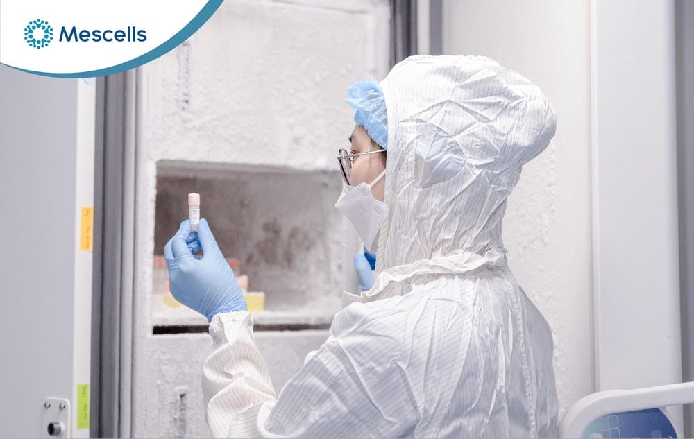
Strict compliance with aseptic procedures, from protective clothing to the handling of cell samples in the culture room
4. Equipment supporting contamination control in the culture room
To put aseptic technique into practice, we need modern physical “fortresses”.
- Biosafety cabinet (BSC): This is the core piece of equipment. A BSC uses HEPA filters to remove 99.97% of particles 0.3 μm in size. Clean air flowing vertically forms an air curtain that prevents dust from the operator falling into the sample.
- CO2 incubator: Many people mistakenly think of this as just a heater. In fact, it is a complex biochemical buffering system. The temperature is held at 37°C, but most important is the 5% CO2 concentration combined with the bicarbonate buffer in the culture medium, which keeps the pH stable at 7.2 – 7.4. Humidity of 95% prevents evaporation of the medium, which could otherwise raise osmotic pressure and shock stem cells.
- Consumables: The use of filtered tips is mandatory to prevent aerosols from the pipette entering the sample. Single-use serological pipettes also completely eliminate the risk of cross-contamination between culture batches.
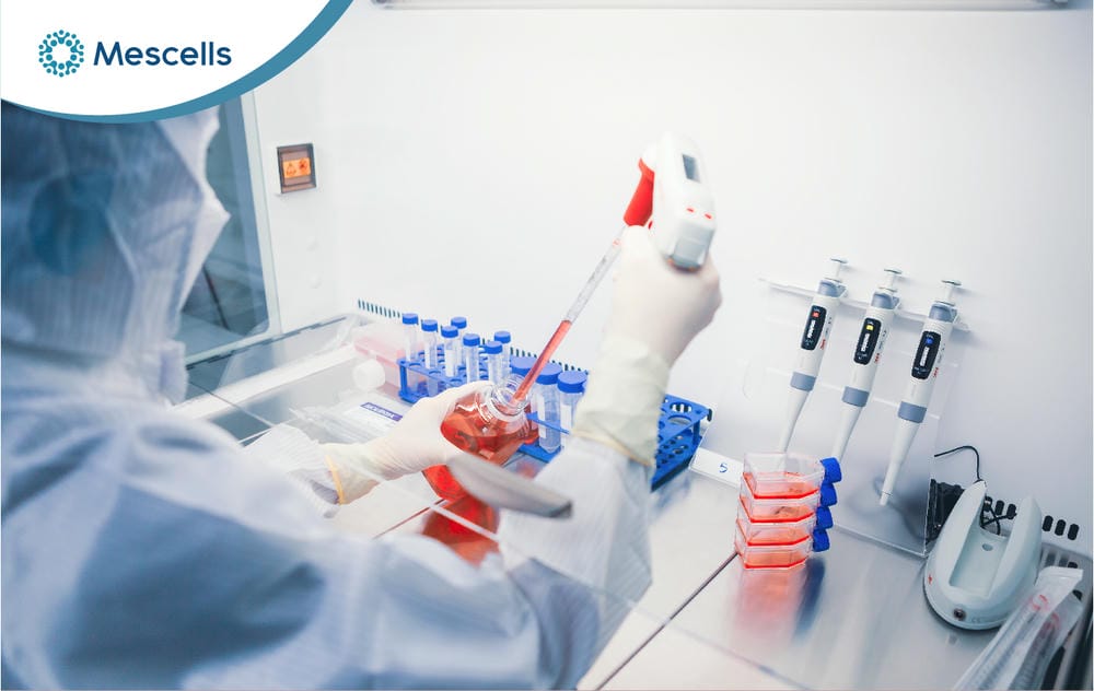
A technician working precisely inside a modern biosafety cabinet
5. The role of contamination control in cell applications
Contamination control in the culture room is one of the key factors determining the quality and safety of cells before they are used in medicine. Especially with stem cells and immune cells, every stage from culture and testing to storage needs to be tightly controlled to limit the risk of contamination and ensure cell quality.
- Vaccine production: Polio, measles and mumps vaccines are grown on clean cells. More recently, adenovirus vectors for Covid-19 vaccines have also required extremely pure host cell lines. If a culture batch is contaminated with bacteria, the entire vaccine lot for millions of people is destroyed, causing enormous damage to global health security.
- Regenerative medicine: After culture, mesenchymal stem cells (MSCs) are delivered directly into the patient’s vein or joint. An unclean cell means introducing a pathogen directly into the body of a patient whose immune system is already weak.
Contamination control is the final barrier between a miraculous therapy and a medical disaster.
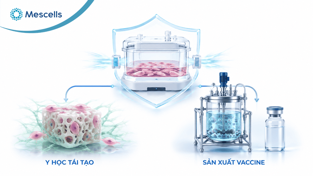
Applications of contamination-controlled cell culture in regenerative medicine and vaccine production
Contamination control in the cell culture room is not just a set of technical steps; it is an expression of respect for science and human life. From understanding the doubling rate of bacteria to mastering 3D bioreactors, every small detail contributes to the quality of the final biomedical product. In the future, technology together with rigorous control procedures will be the foundation for regenerative medicine to develop safely, effectively and sustainably.
MESCELLS | MSC – SPECIALIZED HEALTHCARE SYSTEM FOR REGENERATIVE MEDICINE AND CELL THERAPY
- Hotline: 0866 022 097
- Address: TT21, No. 204 Nguyen Tuan, Thanh Xuan Ward, Hanoi, Vietnam.
- Fanpage: Mescells Institute of Applied Cell Technology Research
REFERENCES:
- Coté, R. J. (2001). Aseptic technique for cell culture. Current Protocols in Cell Biology, Chapter 1: Unit 1.3. https://pubmed.ncbi.nlm.nih.gov/18228291/
- Pipette.com Team. (2026). Cell Culture Contamination: Identifying Different Types. https://pipette.com/blog/cell-culture-contamination
- ECACC Laboratory Handbook. (4th Edition). Cell Culture Protocol 1: Proper Aseptic Technique. Sigma-Aldrich. https://www.sigmaaldrich.com/US/en/technical-documents/protocol/cell-culture-and-cell-culture-analysis/mammalian-cell-culture/aseptic-technique
- Food and Drug Administration (FDA). (2022). Guidance for Industry #253: Current Good Manufacturing Practice for Animal Cells, Tissues, and Cell- and Tissue-Based Products.
Note: The content of this article is compiled from and refers to scientific reports and reputable medical journals around the world. However, this information is for reference only and is not a substitute for diagnosis or specialist medical treatment by a doctor.

Deputy Director, Mescells Institute of Applied Cell Technology Research



 Hotline
 Zalo
Zalo
 Fanpage