 0866 022 097
 institute@mescells.com
Stem Cells in the Joint-Preserving Treatment of Avascular Necrosis of the Femoral Head


Avascular necrosis of the femoral head is common in middle age and is increasingly affecting younger people. If not detected and treated in time, the disease progresses to collapse of the femoral head, making hip replacement unavoidable. Against this background, stem cell therapy in the joint-preserving treatment of avascular necrosis of the femoral head – especially mesenchymal stem cells (MSCs) combined with core decompression surgery – is opening up a new treatment approach that is helping many patients recover. So how is this treatment carried out, and what do studies and clinical trials of the therapy show? Join Mescells to find out in the following article.
Overview of avascular necrosis of the femoral head
1.1. What is avascular necrosis of the femoral head?
Avascular necrosis of the femoral head (osteonecrosis of the femoral head, ONFH), also called aseptic necrosis of the femoral head, is a disease in which bone and bone marrow cells die because of a lack of blood supply to the upper part of the femoral head. Initially, the necrotic area develops zones of reduced bone density and bone defects; this later leads to subchondral fracture and finally to collapse of the femoral head, secondary osteoarthritis and loss of hip joint function, resulting in disability.
Spontaneous necrosis of the femoral head is most common in middle age and affects men more than women. Symptoms include pain in the groin that worsens with walking; pain radiating to the hip, down the thigh and to the knee; limping…

Avascular necrosis of the femoral head develops because of a lack of blood supply to the upper part of the femoral head
1.2. Causes of avascular necrosis of the femoral head
- Prolonged corticosteroid use: The leading cause of non-traumatic necrosis, especially common in patients taking steroids for autoimmune disease, organ transplantation or after COVID-19. Corticosteroids increase pressure within the bone and cause atherosclerosis of the vessels supplying the femoral head.
- Alcohol abuse: Alcohol raises blood lipid levels, blocking the small vessels that supply the femoral head.
- Regular tobacco use: Cigarette smoke generates large amounts of oxidants that damage the lining of blood vessels. Nicotine stimulates vasoconstriction and accelerates the formation of thrombi (blood clots), blocking the very small blood vessels that supply the femoral head.
- Trauma (femoral neck fracture, hip dislocation): Directly interrupts the blood vessels supplying the femoral head, leading to bone necrosis.
- Sickle cell disease, Gaucher disease, gas embolism: Block the microvessels supplying the bone.
- Previous pelvic radiotherapy: Radiotherapy destroys bone-forming cells and the blood vessels that nourish them and damages the endothelium, leaving the femoral head ischemic, shrunken and progressively weakened, and ultimately leading to collapse and bone necrosis.
- Other factors: Autoimmune diseases (such as lupus erythematosus) and chronic conditions such as high blood pressure, diabetes and lipid metabolism disorders… increase the risk of aseptic necrosis of the femoral head, mainly by damaging or blocking the blood vessels that supply it or through the side effects of the drugs used to treat them.
1.3. Current treatments for avascular necrosis of the femoral head
Medical treatment is used for cases without clinical symptoms, with small bone lesions (ideally under 15%), or for patients who cannot undergo surgery. It includes: early removal of risk factors such as corticosteroid abuse and alcohol; bisphosphonates (taken orally weekly or monthly, or given by intravenous infusion once a year if osteoporosis is also present); physical therapy with electromagnetic fields and acoustic waves; and local collagen injection.
Surgery is considered when medical treatment is ineffective. Depending on the degree of necrosis, the doctor will recommend:
- Core decompression of the femoral head for stage I and stage II lesions without collapse of the femoral head. This procedure reduces pressure within the bone, improves blood flow and stimulates the formation of new blood vessels.
- Core decompression combined with bone grafting, using a free bone graft to fill the bone defect or a vascularized bone graft to help rebuild bone structure and improve the blood supply to the femoral head. This approach is usually indicated for stage III necrosis of the femoral head.
- Partial or total hip replacement for necrosis of stage IV or above, where the patient has severe pain, the femoral head has collapsed or become deformed, or severe hip osteoarthritis has developed and conservative measures are no longer effective. Hip replacement gives marked pain relief, maximum functional recovery and improved quality of life.
These methods relieve pain but cannot stop the disease from progressing. After core decompression, as many as 20-40% of patients still deteriorate, with collapse of the femoral head requiring total hip replacement. Mesenchymal stem cells (MSCs), by contrast, can promote new blood vessel growth, regulate immunity, stimulate bone-forming cells and improve hip function, minimizing the risk of femoral head collapse. Combining core decompression with mesenchymal stem cells offers a lasting joint-preserving treatment, with the chance of avoiding surgery and keeping the patient’s natural joint structure.

Hip replacement surgery may be used for stage IV avascular necrosis of the femoral head
2. Using stem cells to treat avascular necrosis of the femoral head
2.1. Stages suitable for stem cell treatment
Stem cell therapy – especially when combined with core decompression – works best in stages I and II, and can still be used in early stage III (IIIA) when the area of collapse is small. A PLOS One review analyzing randomized clinical trials from 2013-2023 confirmed that intervention at an early stage, before the femoral head collapses, is far more effective in preventing disease progression and reducing the rate of joint replacement (according to Robin Novriansyah, 2025):
- Stages I-II: The femoral head is still spherical and has not collapsed, allowing mesenchymal stem cells (MSCs) to attach, promote bone regeneration and form new blood vessels. This is also when stem cell treatment has the highest success rate.
- Stage IIIA: The femoral head has just begun to collapse, with the lesion covering <30% of the head. Stem cells can still be used, but effectiveness declines.
- Stage IIIB and above: The femoral head has collapsed severely and osteoarthritis has developed. This stage is no longer suitable for joint-preserving treatment; the patient needs a hip replacement.
2.2. Types of stem cells used
Mesenchymal stem cells (MSCs) are the most widely studied and applied type in the treatment of femoral head necrosis. The main MSC sources used include:
Bone marrow-derived mesenchymal stem cells (BM-MSC/BMAC): Bone marrow is the most commonly used source of stem cells in the treatment of avascular necrosis of the femoral head. In a suitable environment, bone marrow-derived mesenchymal stem cells can differentiate into cells of the musculoskeletal system, such as trabecular bone, joint cartilage, tendon, etc. Clinical applications of bone marrow-derived mesenchymal stem cells take three main forms: bone marrow concentrate (BMC), cultured cells and bone marrow aspirate, of which BMC is the most widely used (according to Yingxing Xu, 2020).
Peripheral blood mesenchymal stem cells (PBMSCs): Compared with bone marrow-derived mesenchymal stem cells, peripheral blood mesenchymal stem cells have several advantages, such as being more accessible, more cost-effective and carrying a lower risk of contamination with tumor cells. Studies by Song and colleagues in a rabbit model of necrosis induced by liquid nitrogen showed that transplantation of peripheral blood mesenchymal stem cells improved ischemic necrosis of the femoral head by promoting new bone formation (according to H.-J. Song, 2011).
Umbilical cord-derived mesenchymal stem cells (UC-MSC): Umbilical cord-derived mesenchymal stem cells have pronounced stem cell properties and low immunogenicity. They have the advantages of being younger and easier to isolate and expand in vitro, are suitable for allogeneic treatment, and have considerable potential for promoting tissue regeneration.
Adipose-derived mesenchymal stem cells (ADSC): Adipose tissue is another reliable source of stem cells for treating avascular necrosis of the femoral head. Adipose-derived mesenchymal stem cells have several advantages, such as ease of access, higher yield and a differentiation potential similar to that of bone marrow-derived mesenchymal stem cells. Pak and colleagues injected autologous adipose stromal vascular fraction (SVF) percutaneously into the hip joint, which improved patients’ symptoms and promoted bone regeneration in the area of osteonecrosis (according to Jaewoo Pak, 2011).

Types of stem cells used in the joint-preserving treatment of avascular necrosis of the femoral head
2.3. How mesenchymal stem cells work in treating avascular necrosis of the femoral head
Mesenchymal stem cells do not simply “fill in” the area of dead bone. They act through several mechanisms at once:
- Differentiation into bone cells: Mesenchymal stem cells directly generate new bone cells, rebuilding the dead bone structure in the necrotic area. However, the in vivo contribution of this mechanism remains quite limited and controversial.
- Paracrine mechanism: MSCs secrete VEGF (stimulating angiogenesis and forming new blood vessels to supply regenerating bone), BMP-2 and BMP-7 (stimulating bone formation), bFGF (expanding local stem cells) and anti-inflammatory factors, creating a favorable environment for bone regeneration.
- Inhibition of bone cell death: Mesenchymal stem cells secrete molecules that inhibit programmed cell death (apoptosis) of the living bone cells around the necrotic area, thereby preventing the dead zone from spreading further.
- Regulation of inflammation: Chronic inflammation in necrotic bone drives disease progression. Mesenchymal stem cells secrete PGE2, IL-10 and TGF-β, which suppress the inflammatory response, reduce pain and create an ideal environment for regeneration.
2.4. The procedure for treating avascular necrosis of the femoral head with core decompression combined with stem cells
Core decompression (CD) surgery is the technical foundation of stem cell therapy in the joint-preserving treatment of avascular necrosis of the femoral head. The overall procedure usually comprises the following steps:
Step 1: Assessment and preparation (4-6 weeks before surgery)
Magnetic resonance imaging (MRI) and X-rays are performed to assess the location of the lesion and the size of the necrotic area in order to stage the disease accurately.
Step 2: Stem cell harvest
A sample of bone marrow (mainly from the iliac crest) or adipose tissue is harvested using specialized techniques and instruments, then centrifuged to separate and concentrate the mesenchymal stem cells. The extract can be used for local injection or sent to a GMP laboratory for culture and expansion (according to Murat Bozkurt, 2024).
Where umbilical cord tissue-derived mesenchymal stem cells are used, the tissue sample is collected and then culture-expanded in a GMP laboratory.
When the cultured cell samples reach the required number, sample quality is assessed against the following criteria: cell count, viability, CD73/CD90/CD105 markers and sterility.
Step 3: Core decompression surgery and stem cell injection
Under X-ray guidance, the surgeon makes a small incision (about 1-2 cm) near the affected bone. A small guide wire and a special drill (trephine) are used to drill a channel into the dead or diseased bone tissue. This core of bone is removed.
A solution containing mesenchymal stem cells (in saline + 5% human albumin) is then injected directly through the drilled channel into the necrotic area. The whole procedure is carried out under X-ray guidance.
Step 4: Follow-up and recovery
Patients can walk with the support of crutches or a walking frame 1-3 days after surgery. Full weight-bearing on the operated leg should be avoided for 6-8 weeks. MRI and X-rays are taken periodically at 3, 6 and 12 months and annually thereafter to assess progress and the effectiveness of bone regeneration.
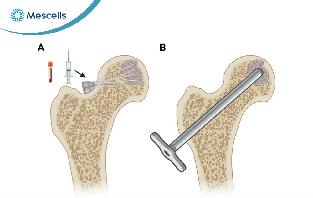
Core decompression combined with stem cells in the treatment of avascular necrosis of the femoral head
3. Studies and clinical trials of stem cell treatment for avascular necrosis of the femoral head worldwide and in Vietnam
A systematic review by Li and colleagues, published in the Journal of Orthopaedic Surgery and Research in August 2023, evaluated the effectiveness of stem cell therapy combined with core decompression in the treatment of avascular necrosis of the femoral head. The study pooled data from 11 randomized controlled trials (RCTs) and 7 retrospective studies, comprising 557 cases treated with core decompression alone and 700 cases treated with core decompression combined with stem cells.
The results showed that the group receiving core decompression combined with stem cells achieved a clearly higher Harris Hip Score (HHS) than the group receiving core decompression alone. The stem cell group also had slower progression of lesions on X-ray imaging and a lower rate of total hip replacement. Notably, no serious complications related to stem cell therapy were recorded (according to Mengyuan Li, 2023).
A comprehensive analysis of studies up to November 2024 (according to Saroj Kundan Bharti, 2025) examined 10 randomized clinical trials with 545 cases and a mean follow-up of 36 months, comparing stem cell therapy with standard treatment in patients with avascular necrosis of the femoral head. It found that stem cell therapy significantly reduced the rate of hip replacement and prevented disease progression. Statistically significant improvements were recorded in the stem cell group compared with the control group on all three scales: HHS, VAS and WOMAC. The findings were assessed using the GRADE approach and confirmed as sufficiently reliable to support recommendations in clinical practice.

A growing number of studies and clinical trials of stem cell treatment for avascular necrosis of the femoral head are being conducted around the world
An open-label Phase I/II clinical trial in Spain followed 8 male patients (mean age 48.38 ± 7.38) with idiopathic avascular necrosis of the femoral head at stage < IIC under the ARCO classification. 40 mL of the patients’ own bone marrow was collected 4 weeks before treatment; mesenchymal stem cells were then cultured to GMP standards and injected at a dose of 0.5-1×10⁶ cells/kg under X-ray guidance.
No adverse effects related to cell injection were recorded. After treatment, pain scores on the Visual Analog Scale (VAS) improved markedly; 50% of patients had improved clinically and 12.5% had improved on X-ray imaging by the end of follow-up. No patient needed hip replacement surgery in the first year after MSC treatment (according to Juan F Blanco, 2023).
The most recent study (according to Oğuzhan Şamil Erciyes, 2025) evaluated 22 patients who underwent core decompression combined with injection of bone marrow aspirate concentrate (BMAC), with a minimum follow-up of 12 months. The Harris Hip Score rose by an average of 23 points (p<0.001), reflecting the clearest clinically meaningful improvement. On X-ray imaging, 100% of stage I lesions regressed to stage 0 (p<0.001). At stage II, 83% of patients remained stable; only 17% continued to progress. The researchers concluded that core decompression combined with BMAC can effectively prevent disease progression at an early stage, preserve the integrity of the hip joint and delay the need for total hip replacement.
In Vietnam, at the 14th Scientific Conference organized by the Ho Chi Minh City Stem Cell Society on 6 December 2025, Nguyen Hai Nam, MSc, representing the Mescells Specialized Healthcare System for Regenerative Medicine and Cell Therapy, gave a report on “Mesenchymal stem cells in the joint-preserving treatment of avascular necrosis of the femoral head”. The report stated that, for patients with early-stage avascular necrosis of the femoral head (stages I and II), a treatment regimen combined with mesenchymal stem cells reduced the joint failure rate to below 10% and cut the risk of joint replacement almost threefold compared with mechanical treatment alone, while maintaining the structure of the femoral head in the long term.
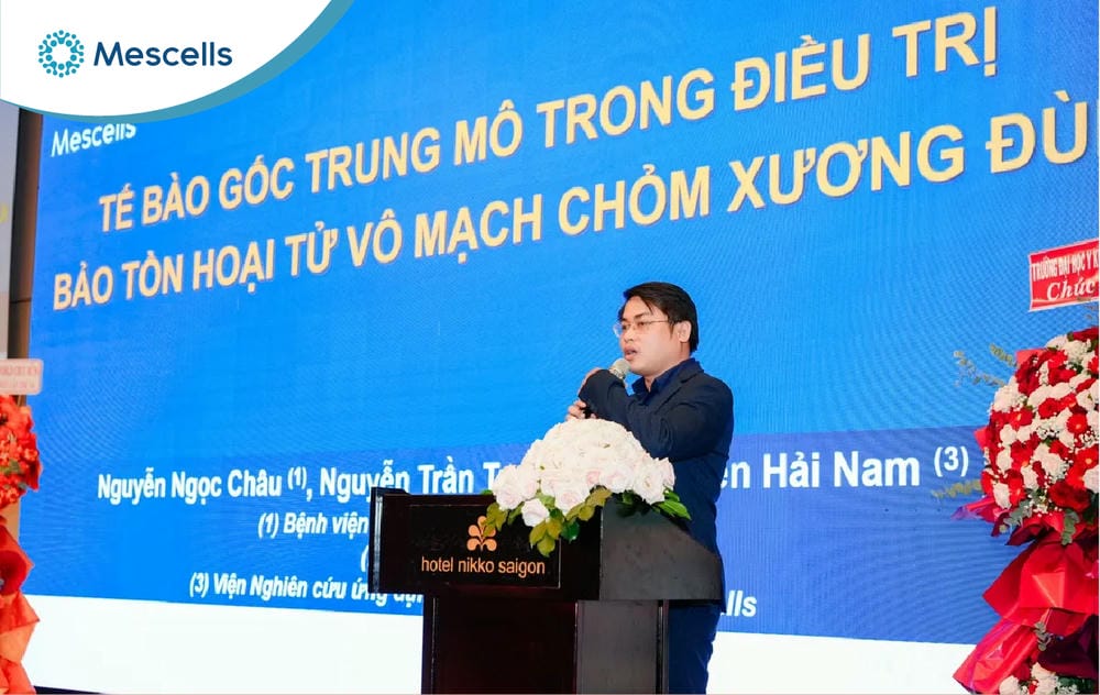
A Mescells representative presenting “Mesenchymal stem cells in the joint-preserving treatment of avascular necrosis of the femoral head” at the 14th Stem Cell Scientific Conference
Applied research using mesenchymal stem cells from bone marrow and adipose tissue has been used to treat cases of avascular necrosis of the femoral head in patients of many different ages. Patients have shown positive improvements both in the structure of the femur on MRI and in their clinical presentation, successfully regaining range of motion and preserving the femur, and avoiding the need for surgery as well as the dangerous complications that might otherwise occur.
Compared with core decompression, which relieves pain effectively but still leaves as many as 20-40% of cases with collapse of the femoral head requiring total hip replacement, the results from mesenchymal stem cells represent a significant advance over the current standard of care.
Stem cells in the joint-preserving treatment of avascular necrosis of the femoral head are an approach increasingly recognized by international science. With the active involvement of many reputable organizations and healthcare systems in research and clinical application in Vietnam, stem cell therapy for femoral head necrosis is gradually being brought into treatment practice, offering hope to patients who want to keep their own hip joint.
MESCELLS | MSC – SPECIALIZED HEALTHCARE SYSTEM FOR REGENERATIVE MEDICINE AND CELL THERAPY
- Hotline: 0866 022 097
- Address: TT21, No. 204 Nguyen Tuan, Thanh Xuan Ward, Hanoi, Vietnam.
- Fanpage: Mescells Institute of Applied Cell Technology Research
REFERENCES:
- Robin Novriansyah. 2025. “Assessing the latest advances in bone marrow stem cell therapy for Avascular Necrosis hip: A comprehensive systematic review, meta-analysis, and meta-regression of randomized controlled trial studies.” PLOS One. https://journals.plos.org/plosone/article?id=10.1371/journal.pone.0297319.
- Yingxing Xu. 2020. “Stem cell therapy for osteonecrosis of femoral head: Opportunities and challenges.” ScienceDirect. https://www.sciencedirect.com/science/article/pii/S2352320420300869.
- Mengyuan Li. 2023. “Stem cell therapy combined with core decompression versus core decompression alone in the treatment of avascular necrosis of the femoral head: a systematic review and meta-analysis.” PMC. https://pmc.ncbi.nlm.nih.gov/articles/PMC10398910/.
Note: The content of this article is compiled from and refers to scientific reports and reputable medical journals around the world. However, this information is for reference only and is not a substitute for diagnosis or specialist medical treatment by a doctor.







 Hotline
 Zalo
Zalo
 Fanpage