 0866 022 097
 institute@mescells.com
What Is Cell Thawing? Principles, Procedure and Factors Affecting Cell Quality


Cell thawing is an important step following the cryopreservation of cells. Performing the technique correctly helps cells maintain their viability, proliferative capacity and biological function. For stem cells in particular, good control of the thawing process has a major bearing on cell quality after recovery. So join Mescells to understand the principles of cell thawing, how the procedure is carried out and how to assess cell quality after thawing.
1. What is cell thawing?
Cell thawing is the process of bringing cells from a frozen state back to conditions suitable for continued culture or use.
During freezing, a cryoprotectant such as DMSO is usually added to cells to limit damage caused by ice crystals. After thawing, DMSO needs to be appropriately diluted or removed, because prolonged exposure can affect the cells.

Retrieving a cell sample from cryogenic storage
2. Principles of the cell thawing process
When thawed, cells undergo rapid changes in temperature, water content and osmotic pressure. If this process is not controlled, the cell membrane and internal structures may be damaged.
The key principles are therefore:
- Warm the cells under defined conditions.
- Limit the time cells are exposed to DMSO after thawing.
- Handle gently to avoid damaging the cells.
- Return the cells quickly to a suitable recovery medium.
No single thawing condition suits all cell types. Parameters need to be optimized for each cell type and specific protocol.
>>> Learn more: Stem cell cryopreservation technology
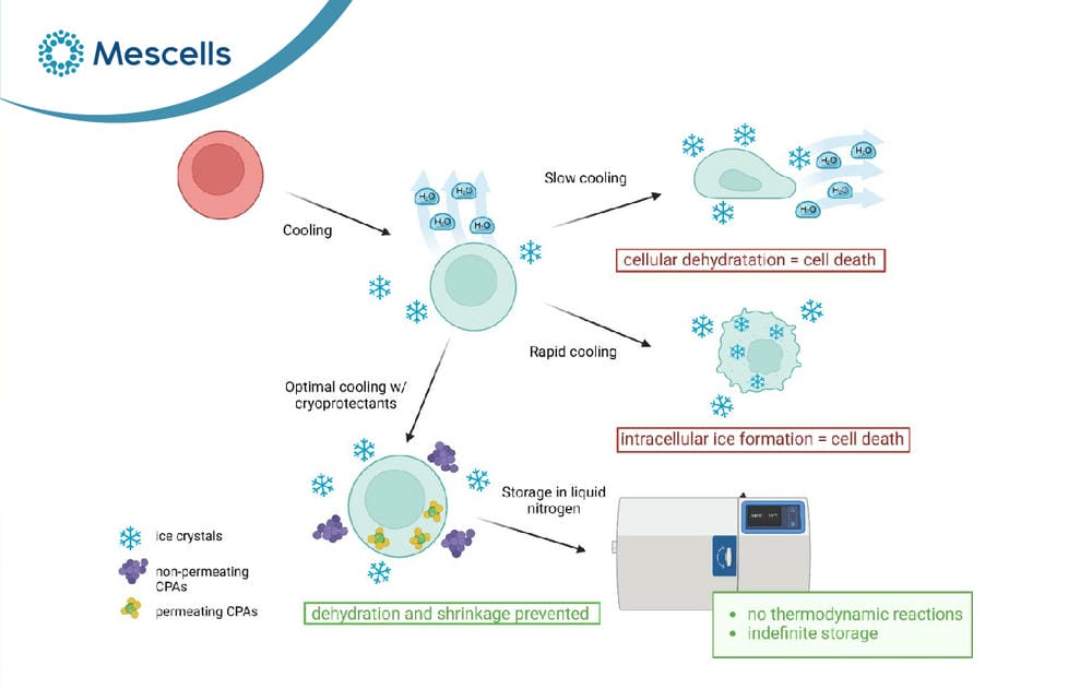
Diagram of the principles of cell freezing, ice crystal formation and the role of cryoprotectants in protecting cells
3. The cell thawing procedure
The actual procedure should be established according to the SOP of each laboratory or manufacturing facility. In principle, however, cell thawing usually comprises the following main steps:
3.1. Step 1: Preparation before thawing
Preparing in advance reduces the time cells spend outside storage conditions and limits unnecessary handling. Before removing the sample from storage, the following should be fully prepared:
- A suitable recovery medium or solution.
- Sterile handling instruments.
- Thawing equipment appropriate to the protocol.
- Tubes or cell containers.
- Tools for assessing cell count and viability. Identification labels and sample traceability records.
3.2. Step 2: Removing the frozen sample from storage
The cell sample is removed from the cryogenic storage system in accordance with sample control procedures. The time the sample is exposed to ambient temperature before thawing begins should be kept to a minimum. Particularly for cell products used in research or clinical applications, control of time and temperature is an important part of the cold chain.
>>> See also: Contamination control in the cell culture room

Placing a frozen cell vial in a water bath for rapid thawing
3.3. Step 3: Warming the sample under established conditions
The frozen sample is warmed according to parameters defined for the cell type and sample volume. In practice, a water bath at around 37°C has been commonly used for many types of frozen cells.
However, controlled thawing systems are attracting growing interest because they give better control over thermal conditions and reduce dependence on manual handling. The goal is for the sample to thaw evenly, without prolonged exposure to unsuitable temperatures.
3.4. Step 4: Diluting or removing the cryoprotectant
After thawing, cells are usually transferred to a suitable recovery medium or solution to dilute or remove DMSO. This is an important step because DMSO protects cells during freezing but can be cytotoxic with prolonged exposure under post-thaw conditions. Post-thaw processing methods may vary depending on the cell type and intended use.
3.5. Step 5: Cell recovery
Once the cryoprotectant has been dealt with, cells are recovered by an appropriate method and placed in culture medium or the corresponding system of use. Excessive mechanical handling should be avoided, because cells after thawing may be more sensitive than cells in stable culture.
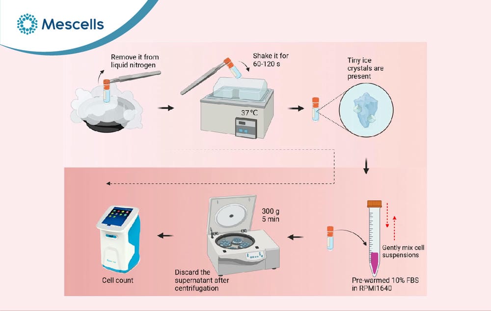
Procedure for thawing and assessing cell viability after cryopreservation
3.6. Step 6: Post-thaw quality assessment
Finally, the cells are checked against appropriate criteria such as:
- Number of cells recovered.
- Cell viability rate.
- Cell morphology.
- Adhesion or proliferation capacity.
- Phenotypic characteristics.
- Specialized function.
- Differentiation capacity for certain types of stem cells.
4. Factors affecting cell thawing outcomes
Post-thaw cell quality can be affected by many factors, the most important being:
- Cell type: each cell type tolerates freezing differently.
- Cryoprotectant: the type and concentration of DMSO can affect cell viability.
- Freezing and thawing rate: affects the formation and recrystallization of ice crystals.
- DMSO exposure time: prolonged exposure after thawing can be toxic to cells.
- Technique: overly vigorous pipetting, mixing or centrifugation can damage cells.
- Sterile conditions: contamination can seriously affect culture results.
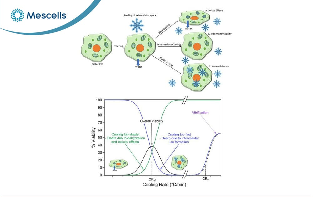
Factors affecting outcomes after cell thawing
5. Assessing cell quality after thawing
A single indicator should not be relied on to conclude whether thawed cells meet quality requirements. A comprehensive assessment needs to consider cell number, viability and function.
- Cell viability rate.
- Number of cells recovered.
- Cell morphology.
- Adhesion and proliferation capacity.
- Phenotypic and functional characteristics, where needed.
High viability is a positive sign but is not enough to confirm that cells are of good quality. For specialized cells or stem cells, activity and biological function also need to be assessed. (Information cited from PubMed – Best practices for cryopreserving, thawing, recovering, and assessing cells)
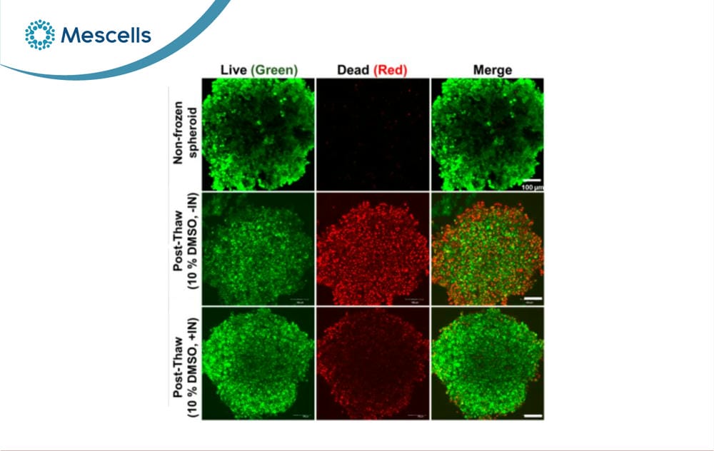
Assessing cell viability and quality after thawing
6. Effects of thawing on stem cell quality
Stem cells are a group of cells that are particularly sensitive to the freeze-thaw process. When assessing stem cell quality after thawing, it is therefore not enough to consider whether the cells are “alive”; one must also consider whether their key properties have been maintained.
- Reduced cell viability.
- Increased cell membrane damage.
- Increased programmed cell death (apoptosis).
- Reduced adhesion or proliferation in the early period after thawing.
- Possible effects on some biological functions of the cells.
Therefore, for stem cells, the assessment should go beyond “are the cells alive or not” to include the cells’ capacity for recovery and their function. (For further information, see the article PubMed – Role of the apoptosis pathway in cryopreservation-induced cell death in mesenchymal stem cells)
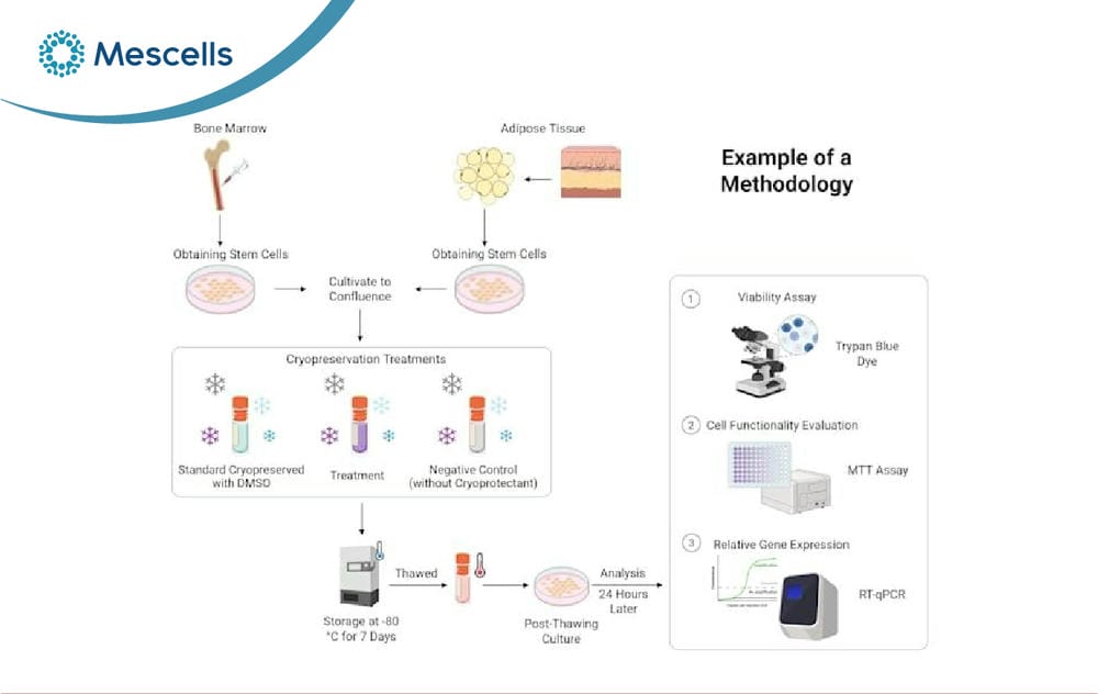
Procedure for culturing and assessing mesenchymal stem cells after freezing
Cell thawing is an important step that determines how well cells recover after cryopreservation. Thawing under the right conditions, reducing DMSO exposure time and handling gently all help limit cell damage. For stem cells, post-thaw assessment needs to consider viability, proliferative capacity and biological function to ensure cell quality.
MESCELLS | MSC – SPECIALIZED HEALTHCARE SYSTEM FOR REGENERATIVE MEDICINE AND CELL THERAPY
- Hotline: 0866 022 097
- Address: TT21, No. 204 Nguyen Tuan, Thanh Xuan Ward, Hanoi, Vietnam.
- Fanpage: Mescells Institute of Applied Cell Technology Research
REFERENCES:
- Baust JM, Campbell LH, Harbell JW. Best practices for cryopreserving, thawing, recovering,andassessingcells2017;53(10):855-871: https://pubmed.ncbi.nlm.nih.gov/29098516/
- Whaley D, Damyar K, Witek RP, Mendoza A, Alexander M, Lakey JRT. Cryopreservation: An Overview of Principles and Cell-Specific Considerations.2021.PubMed:https://pubmed.ncbi.nlm.nih.gov/33757335/
- Heng BC, Ye CP, Liu H, et al. Loss of viability during freeze-thaw of intact and adherent human embryonic stem cells with conventional slow-cooling protocols is predominantly due to apoptosis rather than cellular necrosis. 2006;13(3):433-445. https://pubmed.ncbi.nlm.nih.gov/16374523/
- Pegg DE. Cryopreservation and thawing of cells 2012. https://pubmed.ncbi.nlm.nih.gov/23129152/
Note: The content of this article is compiled from and refers to scientific reports and reputable medical journals around the world. However, this information is for reference only and is not a substitute for diagnosis or specialist medical treatment by a doctor.

Deputy Director, Mescells Institute of Applied Cell Technology Research



 Hotline
 Zalo
Zalo
 Fanpage